EXPLORING WAVEGENETICS AND WAVE IMMUNITY.
THEORETICAL MODELS *.
Gariaev P.P., Kokaya A.A., Leonova-Gariaeva E A., Muldashev E.R., Mikhina I.V.,
Smelov M.V., Tertishnii G.G., Tovmash A.V., Chalkin S.F., Shatrov Y.K., Yagujinski L.S.
* Disclaimer: This translation and English text editing have been done to the best knowledge and understanding of the translator who is not a specialist in the field although having a good understanding of Russian language and advanced level of English complemented with rather good understanding of basic physics and biology. The reader is encouraged to seek specialist opinion in any event. Extensive resources are used to understand the concepts in order for the translation to be s precise and accurate as possible. Where possible Russian abbreviations are to be preferred as they represent the most accurate references. An approach of translating the concepts, not the discrete words, has been employed in a belief that this is the most appropriate form of conveying a thought and/or concept from one language into another; of course a detour from this mode is taken where deemed necessary for the sake of clarity.
All authors have equally contributed to the research in this paper.
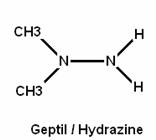
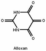
Many researchers have pointed out in their works to the dangers posed by geptil pollution to the environment and that it adversely and detrimentally affects health of livings organisms [http://www.seu.ru/conference/ecoprvo/geptil.htm]. In this present paper a possibility of principled approach to the development of technology is demonstrated, which allows bio-systems to trigger resistance mechanisms to protect the organism from the toxic impact of geptil substance by mean of electromagnetic fields/waves. Theoretical foundations for conducting this kind of research are illustrated on the following website http://www.wavegenetics.jino-net.ru/. Prior to commencing this research/investigation in this direction, we had organised a series of initial model experiments with alloxan. Alloxan is a cytotoxic substance with a dominant detrimental effect on β-cells of pancreas resulting in to Type 1 diabetes.
The earlier experimental works [Gariaev P.P., Kokaya A.A.] have suggested that modulated by pancreas and spleen – modulated wideband electromagnetic radiation (MWER) generated by helium-neon laser influences upon progression of the experimental diabetes in rats. Diabetes is provoked by intro-peritoneal/abdominal injection of alloxan in a dose of 200mg/kg of the mass of the animal.
Irradiating rats with such a frequency resulted in prolonged life expectancy of the animals in the test groups when compared to the control group, leading to normalisation of blood glucose level and, crucially, facilitated pancreas tissue regeneration process.
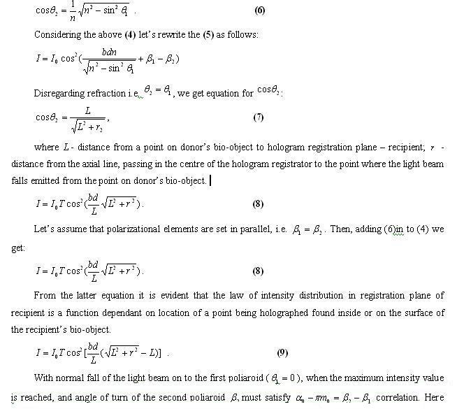
The aim of this present paper is to assess the phenomenon of resistance to alloxan developed in animals having been previously preventively irradiated by MWER. For that purpose a special laser was used which features interconnected complementary orthogonal polarization of the light beam. Generation of the WER (wideband electromagnetic radiation) was conducted in accordance with Fabri-Perot interferometer’s scheme, where the operational laser ray many times passed through the thin freshly-prepared sections/samples of pancreas and spleen from a healthy rat. We believe that the samples specifically and uniquely modulate the laser ray in such a way that this system reveals the following capabilities:
1) reinforce/enhance WER released from discharge interval of He-Ne laser;
2) WER is parametrically connected with the sample-modulated laser ray and as a result it obtains a property of being biologically highly active;
3) biological effect can be observed at relatively long distances from the source of WER;
4) Genetical-metabolic managing/primary information is transmitted from bio-donor to the bio-recipient. The information is carried by MWER (modulated wideband electromagnetic radiation) modulated by the bio-object from the donor
As a donor/bio-structure tissue, probed by the laser beam in the present system, following can be used: living and/or quasi-living organisms, for instance, bacteria, viruses and also living through tissues and organs, metabolites and abiogenic substances.
RESEARCH METHODOLOGY. PHYSICS’ PART
To obtain WER modulated by the bio-structures/samples we utilized developed earlier biotechnology with use of helium-neon laser [Gariaev P.P., Tertishni G.G. 1999]. Helium-Neon laser of 2 MW with 632.8 nm of the wavelength possesses two superposed, orthogonal linear polarized modes of emission, single-frequency in each of them. Laser ray probes/scans DNA samples/bio-structures — newly retrieved tissues of pancreas and spleen of a newborn rat Wistar strain. Semitransparent tissues were deposited onto a a lab glass and then covered that by a second lab glass and this “sandwiched” object was fixed in front of the optical axis of the laser. Adjustment of the glasses with the tissues had been done so as to provide partial reflection of the modulated (by the tissues) laser beam back in to the laser resonator. In this way we ensured a multiple passages of the laser beam through the tissues and the tissues be an optical correlator [Mazur, Grachev, 1985] and therefore affect redistribution of the secondary modes of the laser emissions.
Optical signals were registered and transmitted to an electronic circuit, which governs laser’s generation regimes; frequency stabilization of the coherent radiation is also performed at this stage. In this mode the laser generates, apart from the red light, WER modulated by the tissues – that is – the MWER itself. Distance from the target to the active element of the laser is 11cm.
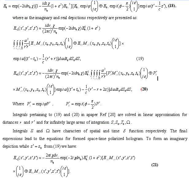
RESEARCH METHODOLOGY. BIOLOGICAL PART
Wistar strain of rats matured to reproductive stage, 5-6 months old were used, with average mass of 180-220 gram. Diabetes for the purposes of this experiment was provoked by way of intro-peritoneal/abdominal injection of alloxan in a dose of 200mg/kg of mass of the animal after 24-hour fasting period with normal/common levels of blood glucose level. The animals were divided into 4 groups:
Group 1 control (n=20) – no WER irradiation; Group 2 (n=20) and Group 3 (n=20) – were subject to preliminary irradiation of WER; Group 4 (n=10) – placebo, where WER was not modulated by DNA samples and the laser ray passed through empty lab glasses without the tissues.
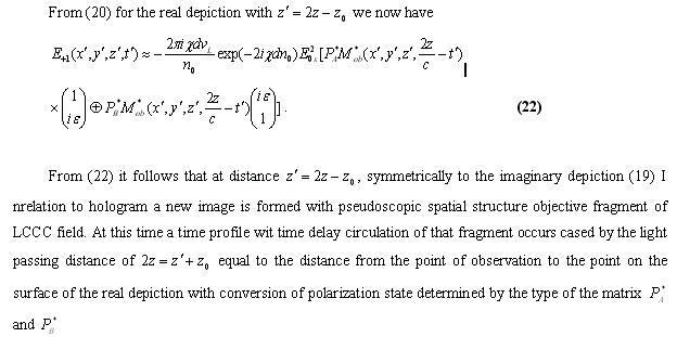
Group 2 rats were placed 20 meters from the source of MWER in the lower ground level of the lab. Alloxan diabetes was provoked one month later from the day of the last irradiation by MWER.
Group 3 and Group 4 were placed 70 cm from the source of MWER. Alloxan diabetes in these groups was triggered one day after the last irradiation by MWER.
Irradiation by MWER of Group 2 and Group 3 was being performed daily for 30 minutes during 4 days scheduled as follows: 10 minutes irradiation by MWER (modulated by samples of pancreas), 10 min irradiation by MWER (modulated by samples of spleen) and 10 minutes irradiation by MWER (modulated by samples of pancreas).
Group 4 – placebo – was being irradiated by WER – not modulated by any samples the laser beam though passing through the empty lab glasses – for 30 minutes daily during 4 days.
Group 1 – control — was not subject to the irradiation by WER nor was it by MWER.
During the experiment we assessed general health condition of the experimental animals, recorded the death date for the animals from the moment of alloxan injection, in all observed groups. The animals of Group 2 and Group 3 were observed for 1.5 months from the day of alloxan injection. In 8 animals from Group 2 and 3 were recorded with the reproductive function still active when the blood glucose level reached its peak value. (3 rats from Group 2 and 5 rats from Group 3).
Measurements of the blood glucose level were performed by glucometer Ascensia Entrust made by Bayer. Measurement range of the glucose level spreads from 2.0 mmol/l up to 30.6 mmol/l. Measurements of glucose level higher than 30.6 mmol/l were marked as HI.
Removal of heart- lung- liver- kidney- spleen- and pancreas- tissues was done in order to perform microscopic description and histological analysis in:
Group 1 – control – on day 3 and 4 after alloxan injection which corresponded to the highest death rate of the animals;
Group 2 and 3 on the day 8 from the alloxan injection, and also on the 42 day of the experiment after assessing reproductive function in male animals.
For histological analysis purposes the tissue had been fixed in 10% neutral formalin, dehydrated in spirits of increasing concentration and then sealed them in paraffin wax. Paraffin sections of 5-7 micro kilometers thin were obtained on microtome Leica SM 2000R, were colored in hematocsilin-eosin and then analysed by microscope Leica DMLS. Video footage was obtained by using CCD-cameras.
Statistical survey of the experiment’s results were conducted by means of using statistical software suits “Stastica 6.0”, MS-Exel” for Windows. Degree of accuracy (p) was determined using Student criterion, employing confidential coefficient and a digit of degree of freedom (1) in accordance with table. Calculations of all mathematical values had been performed in compliance with commonly known formulas on a PC.
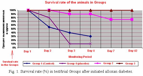
Research in this series of experiments revealed that application of the said dose of alloxan in control and placebo groups developed diabetes complicated by toxic damage of live-crucial organs and systems. This led to low survival rate of the animals in within the groups. On the contrary, in Group 2 and 3 we observed resistance/increased immunity in the animals to the detrimental/destructive effects of alloxan expressed in various degrees.
Group 1 – control – survival rate after alloxan injection of the animals on the day 2 was 55% yet by the day 4 it went down to 30% (Fig.1). Glucose level in Group 1 animals on day 2, 3 and 4 is reliably distinct (p<0.05) from the initial value/level (Table 1). Animals dying in Group 1 (control) at terminal stage were put down by euthanasia (5 rats), organs used for patho-morphological analysis. There was no discrete/spontaneous reduction in glucose level in Group 1 (control) within the period of monitoring. However, there was one rat exhibiting resistance to alloxan and its blood glucose level remained within the limits.
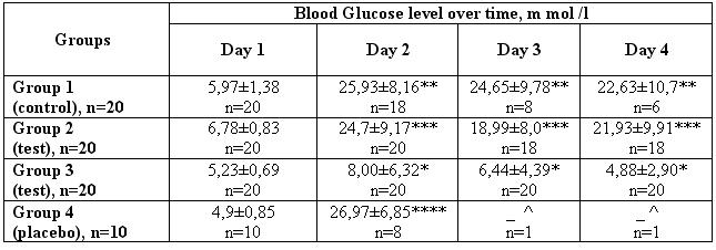
Table 1. Blood Glucose level over time in animals after alloxan injection, dose of 200mg/kg of the mass
* — blood glucose level in Group 3 by the 2nd, 3rd, and 4th day of initiating alloxan diabetes is different (p<0.05) from the level of glucose in blood of the animals in Groups 1 and 2 by the 2nd, 3rd, and 4th day, and is also different (p<0.05) from glucose level in blood from the animals in Group 4 by the 2nd day;
** — blood glucose level in Group 1 by the 2nd, 3rd, 4th day is in fact different (p<0.05) from the initial level;
*** — blood glucose level in Group 2 by the 2nd, 3rd, 4th day is different (p<0.05) from the initial level;
**** — blood glucose level in Group 4 by the 2nd day is different (p<0.05) from the initial level;
^ — in Group 4 by the 3rd and 4th day of observance there was one survived rat.
All animals were injected alloxan – 200mg/kg of an animal mass. Group 1 (control) was not subject to WER or MWER. Group 2 – MWER was conducted; the animals were places 20 meters from the source of emission. Induction of alloxan diabetes was effected one month later after the last radiation by MWER. Group 3 – MWER was conducted, animals were located 70 cm from the source. Induction of alloxan diabetes was done one day after the last radiation by MWER. Group 4 – placebo – was irradiated by WER – not modulated by DNA samples (that is to say that the laser beam was passing through the empty lab glasses). The animals were placed 70 cm from the source of the emission. Alloxan diabetes was induced a day after the treatment.
Animals in Group 4 – placebo — were observed with altered glucose level different from the initial one (p<0.05). 80% of the animal survived by the day 2 and only 10% remained alive by day 4. This was considerably at variance with what we observed in Group 2 and Group 3; Group 1 exhibited lower death rate and survival was at 30%. (Fig. 1)
Preventive irradiation by MWER significantly influences the alloxan diabetes progression in the animals of Group 2 and Group 3 (Figs. 1, 2, 3, Table 1) and is accompanied by safeguarding, cyto-ptotective effect. (Fig. 4). That had been seen in both abovenamed Groups despite the fact that diabetes in Group 2 was induces a month later after the last preventive irradiation/treatment
Survival rate in Group 2 (n=20) reached 90% by the 3rd and 4th day from the moment of alloxan injection (Fig.1) which is significantly distinct from survival data obtained from the control Group (30%) and in the Group 4 (placebo) (10%). Glucose level increase (p<0.05) is observed in Group 2 animals by day 2, 3 and 4 when compared with the initial value (Table 1). By the 4th day 13 animals in Group 2 (65%) the glucose level in their blood was 14.5 mmol/l, whereas 5 animals (25%) from the same group were observed with normal physiological level of glucose (Fig.2)
Glucose level in Group 2 by the 4th day is (p<0.05) different from the initial level. (Table 1). By the 7th day in Group 3 survival rate decreased to 75% (out of 20 animals with hyperglycemia 5 animals died) and remained at the same level up to the end of observation term (1.5 months Fig.1). By the 8th day euthanasia had been conducted to 6 animals from Group 2 and tissues were extracted for patho-morphological analysis. The other 9 animals were observed for a period of 1.5 months. It is important to note that from the day 8 up to 15th animals with hyperglycemia a reduction in glucose level was observed (Fig. 2).
However by the 18th day 4 animals of Group 2 regained the hyperglycemia (more than 30,6 mmol/l) which remained during the entire observation period. The overall health condition of these 4 animals were assessed as satisfactory. Similar results were present during our previous experiments [Gariaev, Kokaya et al, 2007]. The other 5 animals maintained normal level of glucose.
Reproductive functions were noted in 3 animals of Group 2 with hyperglycemia (glucose level over 30.6 mmol/l). These animals gave birth to healthy multiple posterity. After 1.5 months tissues were extracted for the analysis.
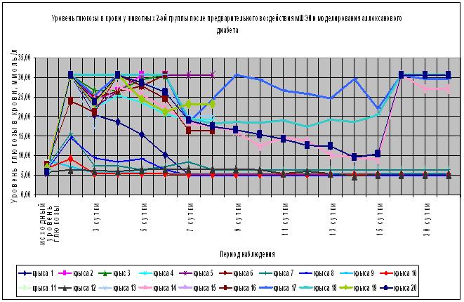
Fig.2 Effects of preventive/preliminary irradiation by MWER on progression of alloxan induced diabetes in Group 2.
Alloxan was injected at 200mg/kg dose one month after the preventive irradiation by [MWER] modulated wideband electromagnetic radiation/field. The irradiation was being performed for 4 days, 30 minutes in each day. Distance from the radiation source – 20 meters. The irradiation schedule: 10 min irradiation time using tissues of the pancreas, then 10 minutes of the spleen tissues, and for 10 minutes – pancreas tissues. The beginning is the day of the alloxan injection. The most manifested effects were observed in Group 3 located at 70 cm distance from the source of the modulated waves. Alloxan diabetes modeling was performed one day after the irradiation by MWER ended (Fig.1,3, Table 1). Every animal in the Group survived. We observed 100% survival rate during the entire period. It is clear that 90% of animals retained their normal physiological glucose level during the 1.5 month of observation, which is clearly different (p<0.05) from the control Group, Group 4 and Group 2.
However 2 animals from Group 3 by the day 6 of the experiment had excessive glucose level increase of 20 mmol/l with subsequent reduction to the normal level. On the 8th day of the experiment6 animals were performed euthanasia for a patho-morphological analysis. Reproductive function could be detected in 5 animals of Group 3. All the animals gave birth to a healthy posterity. 1.5 months later tissues from 6 animals from the same Group were taken for the analysis. Glucose level was at normal levels. Health condition of all the animals of Group 3 was satisfactory.
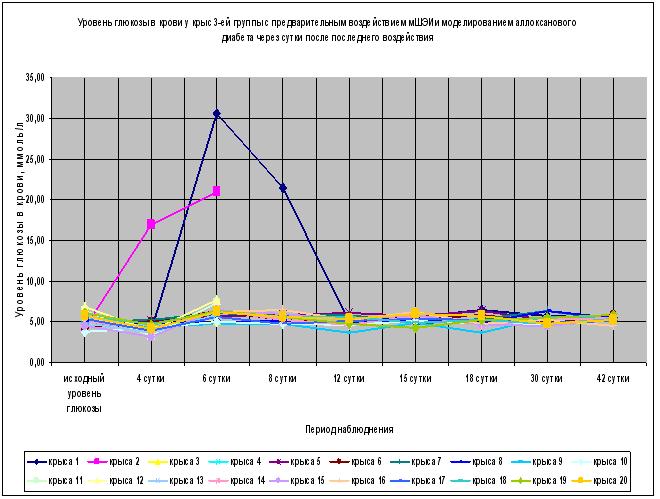
Fig.3 Effects of preventive/preliminary irradiation by MWER on progression of alloxan induced diabetes in Group 3.
Alloxan was injected at 200mg/kg dose one day after the preventive irradiation by [MWER] modulated wideband electromagnetic radiation/field. The irradiation was being performed for 4 days, 30 minutes in each day. Distance from the radiation source – 70 centimeters. The irradiation schedule: 10 min irradiation time using tissues of the pancreas, then 10 minutes of the spleen tissues, and for 10 minutes – pancreas tissues. The beginning is the day of the alloxan injection.
After histological analysis of the pancreas tissues in Groups 1,2 and 3 a number of distinctive features have been observed (Fig.4). Histological picture of the pancreas tissues from animals of control Group was cjaracterised as having clearly visible degenerative alterations of Langer Islands (Fig.4b). The number and size of the islands are reduced, they are of unusual and irregular form. Quantity of β-cells is sharply reduced and in most of them cytoplasmic vacuolation was observed, the nucleus size decrease, chromatin condensation and cariopicnose in some of the cells. Gathering of lymphocyte infiltrate had been revealed around and inside of the islands.
In Group 2 by the 8th day histological situation if pancreas tissues could be described as having damaging processes of various degree: the islands were reduced in size, of irregular form, β-cells reduction, the total portion of insulin apparatus in the islands was significantly reduced. Only a relatively small part of the islands apparatus maintained rather preserved (intact ) structure (Fig. 4с).
Situation in Group 3 by the 8th day after modeling alloxan diabetes was considerably different from the control Group and Group 2. Along with the pathological processes in the tissues of pancreas in question, there was also observed a large quantity of islands, large as well as medium and small sizes with lightened cytoplasm, of normal spherical form, large roun nucleuses containing the core (Fig.4e).
A month and a half later histological situation in Group 2 was also described as having damaged islands’ apparatus (Fig.4d)
As opposed to Group 2 and control Group, Group 3 after 1.5 months had hypertrophy and hyperplasia of the pancreas. Numerous islands of varying size and normal spherical form were noted (Fig.4f). Attention was attracted by the large number of small islands and discreete agglomeration of β-cells, where as large islands contained increased number of β-cells which in turn were located in a close proximity to one another. The structure if the islands and β-cells were not altered, the kernels in the cells were large; round, in which it was possible to locate nucleuses.
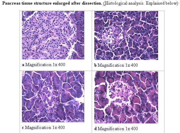
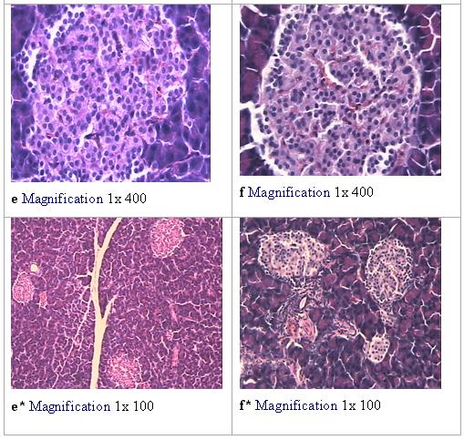
Fig.4 Pancreas tissue structure, Langerhans islands:
a – intact rats:
b – Group 1 (control), after alloxan injection of 200mg/kg dose;
c – Group 2 by the 8th day from the day of alloxan injection of 200mg/kg dose. A month prior to the inducing alloxan diabetes, this Group was preventively treated by the irradiation and was located at about 20 meters distance from the source I the basement of the laboratory.
d – Group 2 after 1.5 months after alloxan injection of 200mg/kg dose.
e – Group 3 by the 8th day from the day of alloxan injection of 200mg/kg dose. One day prior to the alloxan injection this group was also preventively treated by the irradiation being located at 70 centimetres distance from the source of radiation.
f – Group 3 after 1.5 months after alloxan injection of 200mg/kg dose
Enlargement: 1 x 400, 1 x 100. Colouring agents: Haematoxylin and Eosin. DISCUSSING THE RESULTS
It can be stated that a positive effect was reached by using MWER in Groups 2 and 3. Differences in the dynamics of glucose level and survival rates of the animals in these two groups point to an interconnectedness between the length of the irradiation by MWER and modeling the alloxan diabetes. Biological long range defensive effects when irradiating with MEWR which were discovered in our previous experiments [Gariaev, Kokaya et al, 2007] has been confirmed in this series of experiments – this research. The effects is manifested in the glucose level dynamics in Group 2 animals and also in the very fact of the animals surviving in this group, when compared with control Group and placebo Group. In spite of the reliable data (p<0.05) suggesting increase in glucose level in the animals of Group 2 compared to the initial state, and an absence of any reliable differences in the glucose level data obtained from Groups 1 and 2 by the 2nd, 3rd and 4th day of the alloxan injection, survival rate in the Group 2 was high. Evidential hyperglycemia in 20% of the animals in this group did not result in to their death but were remaining I a satisfactory health condition for the entire period of the observation.
Irradiation by the MWER on the animals of Group 3 facilitated development of resistive properties of their immune system to the alloxan triggered diabetes and the glucose level was at the normal physiological lever at all times for the entire period of observation (this is reliably different (p<0.05) from data obtained from Groups 1, 2 and 4). Survival rate in Group 3 was at 100%. Having analyzed the histological data of pancreas from different Groups, it can now be stated that preventive irradiation by MWER in Group 3 resulted not only in cyto-protective effect on the cells of pancreas but also encouraged hypertrophy and hyperplasia processes in it, which, as it seems, were functioning in a compensatory mode. The present experimental findings are in good conformance with the results of our previous experiments conducted earlier. [Gariaev, Kokaya et al, 2007]
In this way, three phenomena of MWER irradiation on the animals with alloxan diabetes were manifestly observed:
Firstly – the factor of survival with contemporaneous and obvious hyperglycemia within prolonged period of observation with preserved reproductive functions in the animals.
Secondly – it was revealed in the previous experiments, and confirmed in the present one that MWER is facilitating pancreas regeneration in the ill animals in situ.
Thirdly – preliminary irradiation by MWER on the animals facilitates development of resistance to alloxan effects.
The recorder effects are related to the fundamental issues of “recording” and transmission of electromagnetic component of genetic information during the post-embryonic growth with wave (frequency) processes taking part in genome and organism as a whole. MWER, parametrically linked with photons (which in turn were modulated by the tissues), appears to be a carrier and transmitter of the information from the bio-donors to the bio-system (bio-organism which precisely and specifically receives this information as strategically superior (that is of higher topology? Of the sort that can manage and govern processes in the recipients organism ) ). Most likely, MWER – performs a function of a frequesncy (wave) trigger, which initiates “awaiting” (stand by) regenerative morphogenetic processes, of which the information is contained in genome of every cell. Quantum mechanisms of MWER effects on embryonic and post-embryonic processes remain to be discovered, thoughsome ideas were expressed in our earlier works. [Garyaev, 1994; Gariaev, 1997; Prangishvili, Gariaev and et al, 2000 (b); Gariaev et al, 2001; Gariaev, 2003] and are being further advances at present. The defensive and cyto-protective action of MWER are promising field of research with rather sound perspectives.
It may be that a specific role in the discovered defensive-protective manifestations plays a so called factor of “weak influence” [Chukova, 2002]. In this regard it can be put forward that the discovered effects bear endoergic character where even weakly absorbed energy of coherent polarized laser beam results in the increase of free Gelmgolc energy which is accumulated in chemical links of tissue metabolites in pancreas and spleen. For instance, atoms of informational macro-molecules (DNA, RNA and proteins), while absorbing light, together with the energy had by a quantum of light they gain the same momentum of quantity of motion, which creates inversed population of nuclear zeeman levels (see Zeeman effect ). Occurs so called chemical polarization of the nucleuses. In this manner, biochemical reactions in preparation (tissues) initiated by the polarized laser light are able to generate electromagnetic radio frequencies (fluctuations). In this situation the preparations (tissues) of the pancreas and the spleen assume a role of specific molecular radio station where every type of molecules has its own frequency (vibration level ) which can be amplified due to stochastic resonance produced by the wideband radiofrequency gas discharge laser.
Based on the obtained experimental data it is proposed to attempt to create a technology allowing development of resistance to toxic effects of Geptil/ hydrazine in animals. We suppose that the resistance to Geptil/ hydrazine and to many other toxic substances can be developed by influencing strategic metabolic vectors most vital of which are – functions of genetic apparatus at quantum level http://www.wavegenetics.jino-net.ru/.
DISCUSSION OF THE EXPERIMENTAL RESULTS.
THEORETICAL MODELING.
The obtained data has wider implications than demonstration of capabilities of possible wave defensive antidote-effect and require theoretical consideration since they relate to strategic (quantum) mechanisms of genetic apparatus functioning of multicell bio-systems. At his stage we propose three formalized hypotheses of the wave processes while “reading or scanning” from bio-structures of donor genetic-metabolic wave information (data), remote addressed transmission of it, introduction of the information in to the bio-system-acceptor and metabolism management using that information.
1. Endogenous polarization-holographic processes in bio-systems
Wave informational scenarios – unfolding in the bio-system itself as well as during scanning by the laser beam at the initial phase – occur at photon level. Let’s consider that level in more details. In our previous researches we presented two- and three-dimensional models of bio-holographic management of building spatial structure of multicell organisms during embryogenesis.At the initial view subject to relatively stationary conditions in bio-systems (final phases of morphogenesis), these models are plausible. However, in living organisms statics and dynamics are paradoxically intertwined.
An adult organism is relatively static spatially on macro scales and significantly changes on this plane only at the stages of deep aging. Along with this that statics are provided for by the internal space-time dynamics of metabolic processes on micro levels of bio-systems organization. This is a case due to the fact that metabolic processes are a mobile aggregate of bio-chemical-bio-physical space-time reformations of the organism’s microstructures. Having considered the non stationary structures of bio-systems, more advanced model is proposed of endogenous informational polarized-holographic managing (governing) processes in multicell organisms, which are realized/effected at genome levels. The model reflects bio-holographic aspect of metabolism as a whole and therefore includes bio-morphogenesis as it specific case. The model utilizes appropriate physics-mathematical formalism for the polarized-holography yet extrapolates it onto probable endogenic analogical processes in the genetic apparatus of multicell organisms.
As a foundation for the model we used our experimental data where we employer special double-polarized He-Ne laser ( = 632,8 nano мeter), having two orthogonal, linked optical modes, of which we have mentioned earlier. When such laser beam of this quantum generator interacts with the bio-tissues, with dynamic holographying on the oncoming beam (bunch) mode on, simultenious acts of recording and scanning of previously unknown information occur, the information about dynamic spinning-fluctuating processes on optical and atomic-molecular levels. Of particular interest can be data obtained about genetic structures and/or living cells. The entire informational structures of an organism, including DNA, RNA and proteins, are optically active, that is to say they are capable of spinning light polarization planes and they are dichroic – difference between absorption of right- and left hand polarized light. Modulations of polarization that correlate with structural-functional condition of any metabolite, present themselves as unique in their capacity storage of information about metabolism and its dynamics and along with it – that is a channel of intercellular photonic bio-symbolic contacts.
Such special processes, in their polarized-holographic version, are apparently inherent to genome workings as a bio-computer. This allows their modeling with use of the aforementioned laser. IT is capable of polarized-holographic recording, scanning, remote transmission and “injection” of wave commanding genetic-metabolic information from one bio-system to another. Besides, such laser performs a conversion of photons probing the bio-system in to wideband electromagnetic spectrum with frequencies from 2 to 0 according to mechanisms of localizing and delocalizing of photons. During this stage, it is apparent, quantum non-local (teleportation) polarized connection across the whole spectrum of frequencies is retained (maintained, saved), including radio waves. Utilization of such laser as reading-transmitting photon-radio-wave system, imitating analogical wave-bio-computer-symbolic non local processes intercellular communications provided with opportunity to carry out remote wave transmission of primary /managing genetic-metabolic information from donor’s bio-system to recipient’s bio-systems (so called addressed transmission ). In the light of this fact it seems rational and vital to attempt presentation of more advanced formalism of bio-symbolic photon-polarized-holographic processes in chromosome apparatus of higher bio-systems, all the more so that the radiowave equivalent of these processes possessed strikingly distinct morphogenetic potencies.
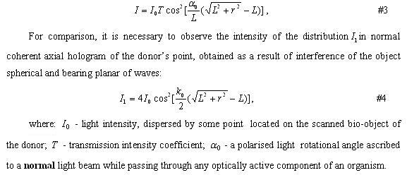
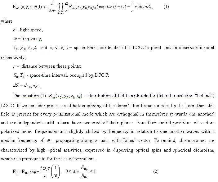
Let’s record vector diffraction Kirchhoff’s integral in paraxial approximation, which describes wave field, for instance photon field, formed by non-stationary bio-system’s fragment. This sort of field may radiate from liquid crystal continuum of chromosome (LCCC) in vivo. Such emission may be expressed by the following:
For the purpose of simplification we will consider that non stationary LCCC is not a function of frequency of translucent light.Both polarisational modes of coheren laser light are depolarized by geno-symbolic acoustic LCCC and are partially elliptically polarized. With this, they can interfere forming specl-structures, and their total intensity is transferred from one mode in to another by means of earlier postulate manner [Prangishvili, Gariaev et al, 2000 (b)].
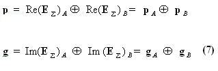
Modified Johns’ vector of every passed orthogonal polarized waves immediately behind the object may be presented in the form of partially coherent orthogonal components of elliptical polarization
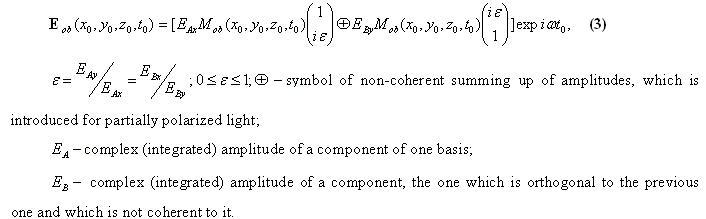
In a bio-system in the composition of LCCC (with only one polarizational component) we use as a hypothetical a carrying wave, which passed, for instance, through infinitely narrow time shitter lock, possessing — figurative characteristic of time transmission (perhaps “gating”). Such a shutter lock completely depolarizes initially polarized wave. The resulting wave, passed behind the gating lock, has continuous spectrum in the whole diapason with evenly distributed spectral density, where as the modified vector of the carrying wave has a form of orthogonal basis of elliptical polarization:
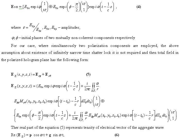
Parameters of the total ellipse p and g are defined via ellipse’s polarization component of every basis A and B, as in Ref [14]
Endogenous biological registration of the summary wave field (5) pertaining to LCCC as a basic element of DNA-wave bio-computer, assumes presence of polarization-sensitive environment in organisms, which is not selective of any particular spectrum across the whole range of active frequencies (similar to the non stationary fragment of biological object, for instance LCCC).
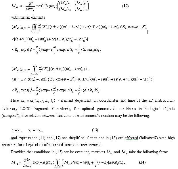
Due to the polarization characteristics of the inducing light in a light-sensitive registering environment of LCCC, photo-anisotropy and photogyrotropy are created (are produced ). To describe the vector photo-response polarized-sensitive environment functions of isotropic , anisotropic — and gyrotropic reactions are introduced, which are constants for every frequency of the active spectrum. Using Johns’ matrixes and rules of their construction for cases of partially polarized inducing irradiation, for the resulting Johns’ matrix we get:
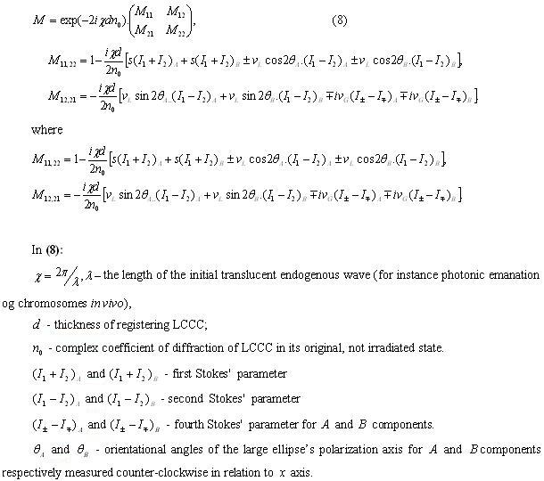
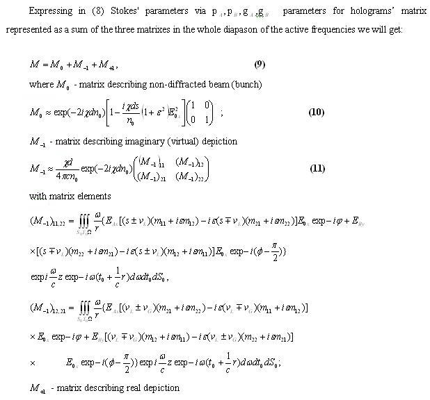
where — LCCC’s hologram fragment size
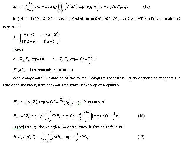
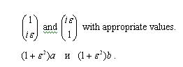
— distance between the point on the hologram surface and the point of observation.Than, successively substituting in (17) expressions for matrixes (10), (14) and (15), let’s define formed by the hologram zero, imaginary and real depictions. It is only now that we can determine, what endogenous or/and exogenous wave for an organism is necessary to utilize as a reconstructing one in order to obtain in the imaginary style regeneration of the required fragment of the wave image of the shaping bio-system. In order to do so it is essential to determine their vectors and corresponding values of matrix. It appears that with precision of up to constant multiplier own vectors of matrix are in essence
It follows that regeneration is performed by a wave identical to the one used while recording by the carrying wave. So, as apparently, in bio-systems at LCCC level recording and reconstruction ensue ither simulteniusly or in accordance with the last equation, then the reconstructed imaginary depiction corresponds to the real one and it is not subject to any distortions. The latter is principallycrucial for preservation of the wave images-vectors of morphogenesis, which compensate physiologic-bio-chemical and mechanical non-stationary stae of the bio-system as a whole and its LCCC in particular. Nonetheless, non-stability of strategic photonic-images of organism’s structures will occur though in term of long lengths of time while aging and its pathological states, for instance in the case of cancerogenesis.
For a passed wave without diffraction the zero depiction has the following appearance:
An analysis of the last correlation shows that with precision up to its multiplier it contains complete reconstruction of space-time structure as well as polarization characteristics of the field of its non- stationary object wave that have passed through, for instance, LCCC. These photo or/and radio wave dynamic structures are, apparently, used by multicellular organisms for their own organization in their own space-time since these structures-images completely retain the true real calibration size without deformations imposed by non-stationary bio-systems and reproduce them in adequate sizes require for a developing or an adult organism. In accordance with the reconstructed wave gradients scanned polarized holograms occurs 4D organization of metabolic “fluxes”/“streams”, cellular architectonics and morphogenetic motions during embryo genesis and also partial regeneration of bio-systems in case of them having been damaged. In other word what occurs is calibration of dynamic potential space-time of bio-system.
Under polarized-holographic-bio-control we imply endogenous or intentional (artificial) modification of recipient’s cell structure and condition as a result of controlling holographic operations from donor’s side. In our case the holographic signal, which was modulated by healthy samples (tissues, cells) of the donor, is transmitted and recorder onto donor’s diseased (ill) cells in form of a hologram. Further, the process of control occurs as follows: in the beginning from the modified cells of the recipient by means of regenerative wave the holographic image of donor’s healthy cells is scanned. It is reconstructed in terahertz wave diapason as a 3D image encompassing every recipient’s cell together with its content.
Mainly, there exist two forms sources of regenerative wave. First form is endogenous. In this instance the processes flow due to innate reserves i.e. “internal” irradiation (emission) of the neighbouring cells. Second form appears to be exogenous reconstruction, when the sources are external emitters. Both forms of sources operate in the recipient’s cells and act simultaneously and continuously, regenerating and complementing the same image of donor’s healthy cells over and over again
According to gradients of intensity of donor’s reconstructed cell images, as if it was a “drawing”, growth and regeneration of recipient’s damaged (ill) cells occur. The cells of the recipient “assume” a role of a photographic film (plate), in which a hologram of the healthy cells is recorded. Processes of the growth and regeneration, flowing analogically to processes of photo-tropism, require certain length of time. In the upshot the recipient’s “sick” cells partially transfer to healthy ranking and partially destroyed. Products of such decomposition are diverted out of the recipient’s organism.
That being so, during the replacement process of recipient’s cells by healthy ones, analogous to those of donor’s, occurs polarized-holographic-control which is in effect germination of the recipient’s damaged cells biomass in to offered to them dynamic holographic form of healthy donor’s cells. As a consequence, while controlling the process, the form and dynamic state (condition) of recipient’s cells is gradually modified under the controlling signal – “pattern”, obtained from the donor.
Needless to say that this form of “pattern” is much more complex than those seen in simple controlling systems. It sets spatial distributions terahertz signal on gradients of which the growth and formation of live cells in recipients occur. Therefore acts of growth and formation of recipient’s cells happen in line with bio-chemical laws, which control their vital functions, and the “pattern” signal sets a program or guide of growth for young cells’ structures and modulation of processes in them.
For more comprehensive description of internal working mechanisms of holographic circular-polarized informational-laser transformation in live organisms it is necessary to have a fair good understanding of basics of polarized-dynamic holographic theory and informational exchange between recipient’s live healthy cells and all other cells included in organs and tissues of the ill organism. When dealing with these issues we utilize de
Transmission of modulated information from donor to recipient occurs by means of rectilinearly propagating longitudinal mutually entwined waves, carrying multilevel modulated information. For the short distance transmissions we can use a term of cellular nucleuses – optical poliaroids (polarizers?), where as for longer distances – a term of quasi lenses (objectives) (here is a reference to some previous papers). Let’s look in to description of such process proffered for purposes of registering color holograms without using lasers [Alexanderov, 1998]. When adapting it to a bio-system, we outline requisite conditions for realizing non-coherent polarized-holographic method of control (management). It ought to be noted that in such systems, having done microscopic studies of the systems, it was discovered long ago that they have microscopic polarisers, i.e. cells’ nucleuses, as well as optically active protein substances, which spin the polarizational plane of emissions that are passed through them. These aspects are long knowng to researchers [Bischof, 1995], however until now this observed phenomenon was neither explained not utilized.
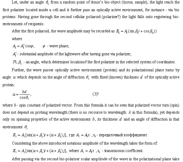
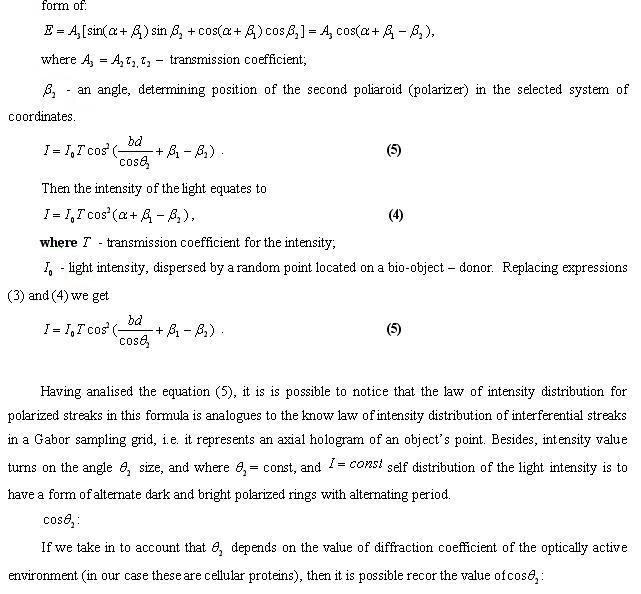
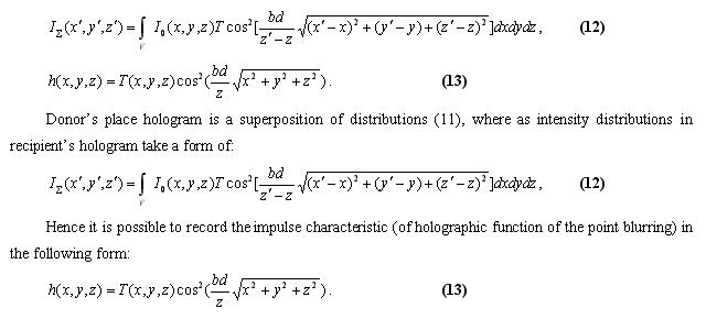
Holographic transmission function can be defined on the basis of Fourier transformation of equation (13). To remind, the wavelength of the probing signal is not included in this formula therefore that wave can be selected from wide range of waves light, electromagnetic and acoustic diapason. The created hologram encompasses whole information about spatial coordinates features of donor’s bio-object being holographed, or about spatial distribution of all donor’s points in relation to holograph registering planes of recipient.
So the resulting solution of the task is, generally speaking, analogical to traditional approach. At the same time the method put forward is fundamentally different from other known interferential methods and possesses certain advantages.
Firstly, instead of some wavelength with its monochromatic features and coherence we use dispercing spinning ability of optically active environments and spatial locally-distributed polarizational filtration. This is sufficient for recording polarized-dynamic hologram of donor (provided that there is motion in donor’s cell in non-coherent wide spectrum irradiation of the recipient)
Secondly, this method allows uncovering the reasons of vibro-resistance when registering and reconstructing holograms without laser sources of light inside bio-systems in terahertz diapason of waves. It efficacy is determined by the value of polarized-optical spinning ability and thickness of the optically active environment . It is known that spinning ability of certain liquid crystals reaches 40000 degrees/mm, which when used in holographic information-laser convertor is sufficient for polarized-holographic transmission of information and accordingly holographic controlling of the biosystem’s structures and processes.
Experimental works on wave interaction in living systems were actively been initiated in 1980s. In the beginning these included researched on interactions between cells [Kirkina et al, 1981; Molchanova, 1985], followed by researches on interaction of living organism [Burlakov et al., 1999]. These works were successfully furthered by A B Budagovski et al. [Budagovski, 1990; Budagovski, Evseeva, 1995; Budagovski et al, 1997; Budagovski et al, 2001]. It was shown that there is a communication and exchange of information of non-chemical (wave, coherent) nature. Such kind of interchange processes, occurring with participation of bio-regulatory signals and occurring without molecular and ion carriers of that information, were labled as processes of remote intercellular interaction (RII) [Budagovski, 2004]. However it seemed incredible that weak electromagnetic cellular signals can produce controlling influences against the background of strong electromagnetic interfering signals of natural and technogenic origins. Nonetheless it appears that with coherent reception light and other electromagnetic non-coherent noises with approximation are zeroed where as those weak coherent and deterministic signals can accumulate [Tertishni et al., 1997, 1998, 2000]
In the last years these works have received the deserved advancements in the Institute of management issues. In particular, utilization of polarized-dynamic holography, which allows formation of little movable polarizational rings. For urposed of transmitting undistorted image of every donor’s point to the remote recipient’s zone, a sensory quasi objective (lenses) were created, and on its basis a holographic device designated for experimental testing of holographic controlling possibilities.